Mr. Jackson
@mrjackson
Mr. Jackson
@mrjackson

Different types of scans you should know about

Have Any Questions?

Please contact us, if you have any queries

Different types of scans you should know about


     When your doctor tells you to go and do a “scan”, what does that mean? There are several types of scans, all for different purposes. Some of them augment the other. This blog will update you on the regularly used scans used to see the organs and parts inside your body.


 Ultrasound is a widely used imaging technique that uses high-frequency sound waves to produce images of internal organs and structures. Ultrasound is particularly useful for imaging pregnancy, kidneys, liver, pelvis, and heart.  When you hear a gynaecologist suggest a scan to see the health of your unborn baby inside the womb, this is the scan that they mean. In fact, for a pregnant woman, an ultrasound scan is one of the safest scanning technique that can be used, as it is free of any harmful radiation.  Ultrasound scanning is less expensive as compared to other scanning techniques and most widely available. 


Another commonly heard word is MRI (Magnetic Resonance Imaging).  The MRI scans use a powerful magnetic field, radio waves and a computer to produce detailed images of internal organs and structures. MRI scans are particularly useful for imaging the brain, spinal cord, joints and soft tissues. 


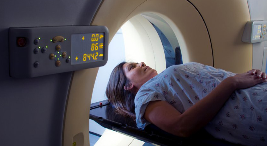
CT scan (Computed Tomography) uses X-rays to produce detailed cross-sectional images of the body. These images are used to identify structural abnormalities, such as tumours, fractures, or abnormal blood vessels.  CT guided procedures can help biopsies from different parts of the body. CT scans are fast, efficient and are also more widely available.  However, CT Scans are not usually advised for pregnancy as it uses X-Rays. 


PET scans (Positron Emission Tomography) use a small amount of radioactive material, called a radiotracer, to produce detailed images of the body’s functions, such as metabolism and blood flow. This allows doctors to identify abnormal cellular activity, such as cancer, before changes are visible on other imaging tests. The radiotracer is usually injected into the patient’s bloodstream, and the patient is then scanned using a special camera that detects the radiation emitted by the tracer. The images produced by the camera are then used to create detailed images of the body’s internal structures. 


PET scan and CT scan are two of the most advanced imaging technologies available today, used for diagnosis and treatment planning of a wide range of diseases. Both technologies have unique capabilities, and when used in combination (called PET CT), they can provide a more comprehensive view of a patient’s condition. The CT scan can show a particular organ and the PET scan can identify where the abnormal metabolic activity is situated in that organ.  This information can be used to do precise needle biopsy from the abnormal site.  The PET CT scan is also used to diagnose the spread of cancer to other parts of the body (secondaries/metastacis).   


Nuclear Medicine 

Another advanced imaging technology is Nuclear Medicine, which uses small amounts of radioactive material to produce images of internal organs and structures, using Gamma camera. Nuclear medicine scans are particularly useful for imaging the bones, thyroid, heart and kidneys. 


New Developments in this area 

In addition to these advanced imaging technologies, there are also new and emerging technologies that are being developed to improve the diagnosis and treatment of diseases. One such technology is Molecular Imaging, which uses small amounts of radioactive material to produce images of specific molecules in the body. This allows doctors to identify early-stage cancer and track the progression of the disease. 


Another emerging technology is Optical Imaging, which uses light to produce images of internal organs and structures. This technology is still in the early stages of development, but it has the potential to revolutionize the way we diagnose and treat diseases in the future. 


Ambady Scan Centre, the best scanning centre in Thrissur has invested in the latest available technology in ultrasound scanning ever since its inception in 1996. At that time, colour doppler was the latest invention to reach the Indian shores, but now Ambady Scan Centre has come a long way and has all the latest technology making it the best pregnancy scanning centre in Thrissur. With more than 25 years of experience and the latest technology, Ambady Scan Centre can claim to be the best ultrasound scanning centre in Thrissur. 


Tags :
Blog
Share This :

Leave a comment

Your email address will not be published. Required fields are marked *