
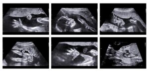
This is the most common type of ultrasound scan
An anomaly scan is a detailed ultrasound scan usually done during 18 to 20 weeks of pregnancy mainly to detect the anomalies of the fetus, its organs, and maternal pelvic organs. This scan increases the prenatal detection of fetal anomalies like cleft lip, club foot, spinal deformity, heart problems, etc. Some anomalies are harmful to the mother and baby and hence, if found earlier, can be treated. That said, while some anomalies can be completely treated, some cannot be. An anomaly scan is generally safe for the baby as it uses sound waves that are not as harmful as a CT scan or X-ray. The scan does not require any preparation, you can have your food. No need for a full bladder for the scan during this period. Here are 5 important points that you should know about fetal anomaly scans.
1. What is examined in an Anomaly scan?
· Thorough examination of baby’s brain, face, heart, stomach, spine, kidneys, hands, arms, legs, and feet.
· Measurements are taken mainly of the head, abdomen, and thigh bones.
· Spine is studied by length and cross-section and whether the skin covers the spine at the back
· Heart is checked for chambers, size, and the major vessels
· The abdominal wall is examined for the placement of internal organs, placenta, and amniotic fluid
· Presence of both kidneys and urinary bladder
· The number of blood vessels in the umbilical cord
· Placenta and its position (to exclude low-lying placenta)
2. What all physical defects are detected at a 20 weeks anomaly scan?
- Spine defects
- Improper formation of the skull bones
- Increased fluid accumulation in the brain
- Heart defects.
- Defects in the diaphragm causing a diaphragmatic hernia
- Defects in the abdominal wall causing intestines to come out
- Missing or malformed kidney
- Missing or malformed bones
- Malformed arms and legs
3. Are there any limitations involved in the scan?
YES, there are some limitations to the scan. While most of the anomalies can be identified. It is not 100% like any other mortality. Some defects evolve only very late in pregnancy and hence, cannot be detected during the 18- 20-weeks window. Several factors like obesity, the position of the baby, twins or triplets, and overlapping of babies can make it difficult to properly visualize certain anomalies. Some conditions like soft tissue fusion of fingers/toes, absence of anal opening or auditory opening cannot be detected by the scan. Similarly, a cleft lip can be detected but further investigation inside the baby’s mouth is limited. An anomaly scan at 18- 20 weeks cannot 100% guarantee an absolutely healthy baby at 40 weeks because many developmental activities are occurring during this period. Still, as a generalization, the majority of the anomalies are detected at 18 – 20 weeks in experience hands with an advanced ultrasound machine
4. Can I identify the gender of the baby?
In India, it is illegal to disclose the gender of the baby during pregnancy through an
anomaly scan. Since gender detection is illegal, the study of genital organs is also
prohibited hence any abnormality in the genital area is most often known only after the
baby is born. At Ambady Scan Centre, we DO NOT perform gender detection of the fetus.
5. Should the baby be aborted in case any abnormality is detected?
Depending on the type of abnormalities, if detected at an early stage of pregnancy, it can be treated. An option for abortion is only suggested in case of dangerous abnormalities with no treatment after birth and appears to be harmful to the mother. The doctors can only suggest and advise on the condition and the relevant remedy, the parents are the decision- makers and it is their decision at the end of the day that counts.
Sounds are a newer form of imaging and are usually performed to find out more about any abnormalities the baby may have. 3D ultrasounds also work well for a woman who is carrying multiple babies, allowing them to clearly see each baby individually.
3D ultrasounds can be done at any time during pregnancy but are mostly performed after 18 weeks. It is useful in determining the baby’s facial features, including the cheeks, nose, mouth, and chin. As the child develops, other body parts can also be seen, as well as internal organs. Unlike 2D ultrasounds, this scan is not that common and requires special probes.
The 3D ultrasound is also a more time-consuming procedure. This technology is particularly useful for families who need to cope with difficult circumstances. This is because the 3D ultrasound can reveal if something is wrong with the baby’s development even before the birth as a photographic image, hence parents can understand better the fetal abnormality.However, 3D ultrasound is not meant to replace a 2D ultrasound.
